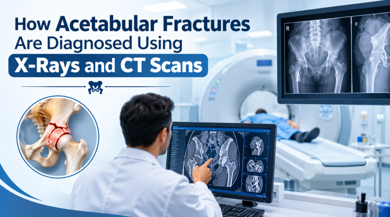
How Acetabular Fractures Are Diagnosed Using X-Rays and CT Scans
An acetabular fracture is a break in the acetabulum, the cup-shaped part of the pelvis that holds the head of the thigh bone (femur). This socket forms an important part of the hip joint and supports body weight during walking, standing, and other daily activities. When the acetabulum is fractured, it can affect hip stability, movement, and long-term joint health.
Acetabular fractures commonly occur after road traffic accidents, falls from a height, or other high-impact injuries. In older adults, weakened bones may also fracture following a fall. Because the acetabulum has a complex three-dimensional structure, accurate diagnosis is essential for understanding the injury and selecting the appropriate treatment.
Doctors generally use physical examinations and imaging investigations, particularly X-rays and computed tomography (CT) scans, to assess these fractures. Each test provides different information about the bone, joint alignment, and fracture severity. Patients consulting an Acetabular Fracture specialist in Jaipur can discuss their imaging results and understand which treatment options may be suitable for their condition.
What Is an Acetabular Fracture?
The acetabulum is the socket in the pelvis that holds the rounded head of the femur. Together, these structures form the hip joint, allowing the leg to move in different directions while supporting the body’s weight.
When the acetabulum breaks, the fracture may affect one or more sections of the socket. Some fractures remain stable, while others cause bone fragments to shift from their normal positions. The severity depends on the fracture pattern, the amount of displacement, damage to the joint surface, and any associated injuries.
Common symptoms include:
- Pain around the hip, groin, or pelvis
- Difficulty standing or putting weight on the affected leg
- Swelling and bruising around the hip
- Restricted hip movement
- Pain that increases when moving the affected leg
- Difficulty walking after an accident or fall
These symptoms require medical assessment, particularly following significant trauma. Severe pain, suspected pelvic injury, or an inability to bear weight should be assessed urgently. Patients should not attempt to walk on a potentially fractured hip before receiving medical advice.
Why Is Accurate Diagnosis Important?
Accurate diagnosis helps doctors determine the location of the fracture, whether the bone fragments have moved, and how the injury affects the hip joint. It also helps identify associated injuries that may not be obvious during an initial examination.
An acetabular fracture that heals with significant joint-surface irregularity or poor alignment may increase the risk of chronic pain, restricted movement, and post-traumatic arthritis. The risk varies according to the severity of the fracture and the success of treatment.
During the initial assessment, the doctor may ask how the injury occurred, review the patient’s symptoms, and examine the hip and pelvis. Circulation and sensation in the affected leg may also be checked. In serious accidents, the medical team may investigate other injuries involving the pelvis, abdomen, blood vessels, or nerves.
Imaging tests are an important part of this process. X-rays commonly provide the initial assessment, while CT scans offer a more detailed view when the fracture is complex or additional information is required.
How X-Rays Help Diagnose Acetabular Fractures
X-rays are often the first imaging investigation when a doctor suspects a fracture around the hip or pelvis. They use a small amount of radiation to produce images of the bones, helping doctors identify many fractures and evaluate the general alignment of the hip joint.
1. Identifying the Fracture
An X-ray may reveal a break in the acetabulum, an irregular bone outline, or an abnormal appearance of the hip socket. It can also help determine whether the head of the femur remains positioned correctly within the socket.
2. Checking Bone Alignment
Doctors assess whether the fractured bone sections remain in their normal positions. If fragments have shifted, the X-ray may show changes in the shape or alignment of the hip socket.
This information helps the doctor determine whether further imaging is needed to evaluate the injury more thoroughly.
3. Examining the Hip Joint
X-rays can reveal changes in the relationship between the femoral head and the acetabulum. They may also identify associated injuries, such as certain pelvic fractures or hip dislocations.
4. Taking Different X-Ray Views
Depending on the suspected injury, doctors may request multiple X-ray views. Different angles help reveal fracture lines that may be difficult to identify in a single image. Special pelvic views may also be used to assess the acetabulum.
However, X-rays have limitations. The acetabulum has a complex structure, and overlapping bones can obscure certain fracture lines. As a result, an X-ray may not show the full extent of the injury. A CT scan may be recommended if the fracture requires more detailed assessment.
Why Are CT Scans Used for Acetabular Fractures?
A computed tomography scan, commonly called a CT scan, produces detailed cross-sectional images using X-rays taken from different angles. A computer combines these images to provide a clearer view of the bone’s structure.
CT imaging is particularly useful for acetabular fractures because the hip socket has several sections and surfaces that may be difficult to assess using standard X-rays alone.
1. Understanding the Exact Fracture Pattern
A CT scan helps doctors identify which parts of the acetabulum are fractured. It may show whether the injury affects the front portion, back portion, or multiple sections of the socket.
This information helps the treating team classify the fracture and understand its complexity.
2. Detecting Displaced Bone Fragments
Some fractures cause pieces of bone to move away from their normal positions. CT scans provide detailed views of the location, direction, and extent of this displacement.
The position of the fragments is important because it can affect hip stability, joint alignment, and the choice of treatment.
3. Assessing the Joint Surface
The acetabulum has a smooth surface that allows the femoral head to move within the hip joint. If a fracture disrupts this surface, normal joint movement may be affected.
A CT scan helps doctors assess the extent of the injury to the joint surface and identify bone fragments that may influence treatment decisions.
4. Planning Surgical Treatment
When surgery is being considered, CT images help the surgeon understand the fracture’s anatomy in greater detail. This information can assist in planning how the bone fragments may be repositioned and stabilised.
Surgery may be considered when there is significant displacement, an unstable hip joint, or disruption of the joint surface. However, not every acetabular fracture requires surgery. The decision depends on the fracture pattern, joint stability, the patient’s overall health, and other clinical findings.
X-Ray vs. CT Scan: What Is the Difference?
| Feature | X-ray | CT scan |
| Main purpose | Initial assessment | Detailed fracture evaluation |
| Image detail | General view of the bones | Detailed cross-sectional images |
| Fracture pattern | Identifies many fractures | Helps define complex fracture patterns |
| Bone displacement | Shows general alignment | Shows fragment positions in greater detail |
| Treatment planning | Useful for initial assessment and follow-up | Often valuable for complex fractures and surgical planning |
Both investigations have important roles. X-rays are often sufficient to identify many fractures, but CT scans may be needed when doctors require more detailed information about the injury.
What Happens During the Diagnostic Process?
The diagnostic process depends on the severity of the injury and the patient’s overall condition. It generally involves the following steps.
Step 1: Medical examination
The doctor asks about the accident or fall, reviews symptoms, and examines the hip and pelvis. Circulation and sensation in the affected leg may also be assessed.
Step 2: Initial X-rays
Pelvic and hip X-rays are commonly taken to look for fractures, dislocations, and changes in joint alignment.
Step 3: CT scan when required
If an acetabular fracture is identified or strongly suspected, a CT scan may be recommended to define the fracture pattern, displacement, and involvement of the joint surface.
Step 4: Reviewing the findings
The doctor considers the imaging results alongside the physical examination, associated injuries, age, general health, and mobility requirements.
Step 5: Developing a treatment plan
Based on the findings, treatment may involve activity restrictions, pain management, mobility support, monitoring, or surgery. Follow-up imaging may be recommended to assess healing and joint alignment.
In selected cases, an MRI may help investigate suspected soft-tissue injuries or other problems that are not adequately explained by X-ray or CT findings.
Treatment Options After Diagnosis
Treatment depends on the fracture’s stability, the amount of displacement, the condition of the hip joint, and the patient’s overall health.
Non-Surgical Treatment
Some fractures can be managed without surgery when the hip joint remains sufficiently stable and the bone fragments are acceptably aligned. Treatment may include:
- Pain relief prescribed by the doctor
- Restrictions on putting weight on the affected leg
- Crutches, a walker, or other mobility support
- Regular medical examinations and follow-up imaging
- Rehabilitation exercises when appropriate
Patients should follow medical instructions about weight-bearing carefully. Putting excessive weight on the injured hip too early may interfere with recovery.
Surgical Treatment
Surgery may be recommended when the fracture causes significant displacement, compromises the joint surface, or makes the hip unstable. One possible procedure is open reduction and internal fixation (ORIF), in which the surgeon repositions the bone fragments and secures them using plates and screws.
The aim is to restore joint alignment, improve stability, and support bone healing. The exact procedure depends on the fracture pattern and the patient’s medical condition.
An Acetabular Fracture specialist can explain the available treatment options, potential complications, and expected recovery process based on the patient’s individual diagnosis.
Choosing an Orthopaedic Specialist in Jaipur
Acetabular fractures can be complex injuries, so patients may benefit from consulting an orthopaedic surgeon experienced in pelvic and acetabular trauma. When choosing an Acetabular Fracture specialist in Jaipur, consider the doctor’s qualifications, experience managing complex fractures, access to suitable imaging facilities, surgical expertise where required, and rehabilitation support.
Patients searching for the best ortho surgeon in Jaipur should evaluate relevant experience, hospital facilities, communication, and the proposed treatment plan rather than relying on a title alone.
A specialist can explain the imaging findings, discuss whether surgery is necessary, and recommend appropriate follow-up care. Recovery may take several months, depending on the severity of the fracture and the treatment provided. Physiotherapy is introduced according to the fracture’s stability and the treating surgeon’s instructions.
Frequently Asked Questions
1. Can an acetabular fracture be missed on an X-ray?
Yes. Some fractures are difficult to identify because of the acetabulum’s complex shape and overlapping bones. If symptoms and examination findings suggest a fracture, the doctor may recommend a CT scan or another investigation.
2. Is a CT scan necessary for every acetabular fracture?
Not always. The need for CT imaging depends on the X-ray findings, the suspected injury, and whether more detailed information is required for treatment planning. CT scans are frequently useful for complex fractures.
3. Can a CT scan determine whether surgery is needed?
A CT scan provides important information about the fracture pattern, displacement, and joint surface. However, the decision to operate also depends on hip stability, associated injuries, the patient’s health, and clinical examination findings.
4. How long does an acetabular fracture take to heal?
Bone healing often takes around 8–12 weeks, although recovery may take longer depending on the fracture’s severity, treatment, age, and general health. Returning to normal activities may require additional rehabilitation.
5. Can someone walk after an acetabular fracture?
Some patients cannot walk because of pain or instability, while others may be allowed limited or protected weight-bearing under medical supervision. Patients should not attempt to walk on a suspected fracture before medical assessment.
6. When should I consult an acetabular fracture specialist?
Seek urgent medical assessment after significant trauma, severe hip or groin pain, difficulty bearing weight, or a suspected pelvic fracture. After initial emergency care, an Acetabular Fracture specialist can help assess the injury and explain the next steps in treatment.
Conclusion
X-rays and CT scans play complementary roles in diagnosing acetabular fractures. X-rays provide an initial view of the hip joint and identify many fractures, while CT scans reveal the fracture pattern, bone displacement, and joint surface in greater detail. Together with a physical examination, these investigations help doctors select an appropriate treatment plan.
Early assessment, accurate imaging, careful follow-up, and rehabilitation when appropriate can support recovery and help protect hip joint function. If an acetabular fracture is suspected, seek prompt medical care and discuss the diagnosis and available treatment options with a qualified orthopaedic specialist.